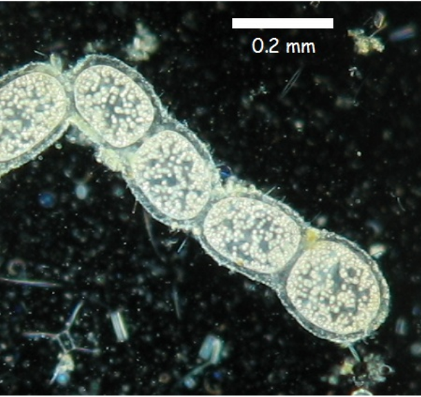
Thiomargarita namibiensis was first discovered in 1997 in marine sediments off the continental shelf of Namibia is known as the world’s largest bacterium1. This bacterium belongs to the Class Gamma-proteobacteria and has a diameter of 0.1–0.3 mm (100–300 μm). However, some reach up to a size of 0.75 mm (750 μm) and is large enough to be visible to the naked eye. The difference in genomic size between T. namibiensis and E. coli is comparable to that of blue whales and newborn mice. Thiomargarita means Sulfur Pearl, Namibiensis means Namibia, and together, it means “Sulfur Pearl of Namibia”. They contain sulfur granules that scatter incident light which makes cells glow white. Just as the division of many cocci, its cell division tends to occur along a single axis, and forms a chain-like pearl string because it is wrapped by a mucous sheath that inspired the name, Namibia’s sulfur pearl. Although it was discovered recently, T. namibiensis is a dominant organism in the seabed of the Namibian continental shelf by representing a biomass of 180g per square meter and accounting for 0.8% of the sediment volume.

Heide N. Schulz and his colleagues from the Max Planck Institute for Marine Microbiology discovered this giant T. namibiensis bacterium while searching for other sulfide-eating bacteria, Thioploca and Beggiatoa. T. namibiensis shares many features with these relatives. They contain vacuoles in unusually large cells, and the cytoplasm is confined to a thin layer of 1-2 μm surrounding the vacuoles. Because the thickness of the activated cytoplasm is not much different from that of normal-sized bacteria, these giant cells can diffuse nutrients as other prokaryotes without special transporting systems. They also store numerous sulfur inclusions that can be used in sulfur-lacking environments. In addition, Osvaldo Ulloa and his colleagues confirmed that their vacuoles contained very high concentrations of nitrate (>0.1 M) in 19953. A notable difference between Thiomargarita, unlike Beggiatoa or Thioploca, is that Thiomargarita does not form filaments. Its cells are wrapped in a mucous-like layer that is attached to each other after cell division, and no motility is observed with the sticky sheath.
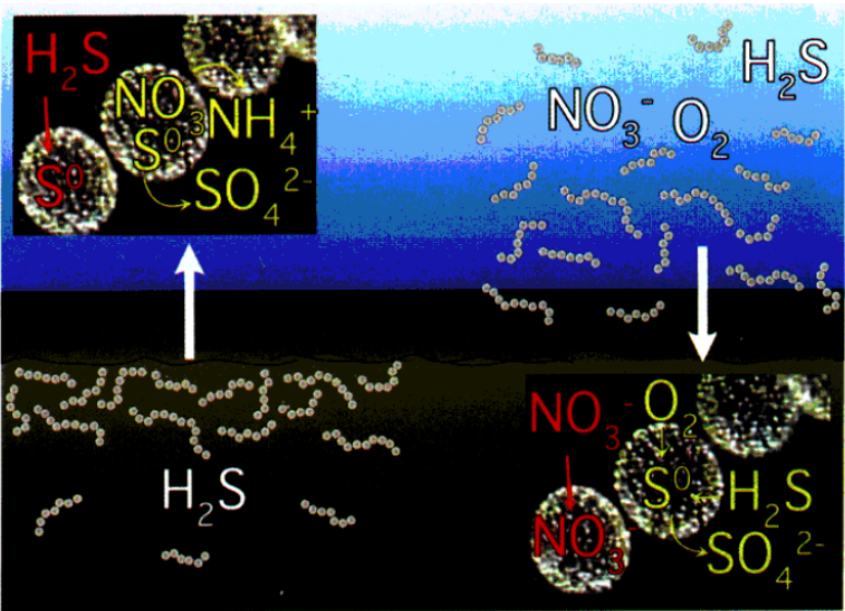
T. namibiensis is a lithotroph and their relatives. Thioploca and Beggiatoa all use sulfide and nitrate (instead of oxygen) for an energy source and an electron acceptor for life activities respectively. Namibia coastal sediments are highly fluid and rich in diatom shells which increase the sulfide concentration by up to 10mM. Nitrate is dissolved in the water near the sediment in a low concentration. Thioploca, with its filament-based mobility, can obtain both sulfides in sedimentary layers and nitrates in the water on site. On the other hand, T. namibiensis that is trapped in sediments does not have difficulty obtaining sulfides, but it must survive in the environment that is not exposed to nitrates because of its immobility. However, sedimentary layers can sometimes rise near the surface due to activities such as methane eruptions, which allows T. namibiensis to store lots of nitrate. The enormous size of Thiomargarita can be interpreted as an adaptation for surviving long periods in the environment with no exposure to nitrates, which act as electron acceptors. If Thioploca obtains spatially separated nutrients, Thiomargarita uses time differences to obtain nutrients.

After the discovery of T. namibiensis, very similar bacteria were found in the Gulf of Mexico in 20055. The cells have the characteristics of Thiomargarita, such as having spherical cell morphology, large vacuoles, and thin cytoplasm with sulfur inclusions. However, they were observed to be divided into 2, 4, 8, or 16 cell clusters without dividing in a single axis to form a “pearl chain”, which showed a clear morphological difference. In addition, another morphological variety of Thiomargarita was confirmed in 20116. Researchers exploring the ecology of methane seeps (a type of symbiotic group) along Costa Rica’s Pacific Ocean (depth 990-1600m) have recognized the familiar form of Thiomargarita in the sediment; they form microbial biofilms to cover the surface. They thickly cover some natural carbonate rocks derived from methane and byssal threads that attach mussels to the substrate. Often, they have been observed to reproduce in the form of budding. Adaption of Thiomargarita in an undersea environment which is completely different from the land allowed us to see how diverse and unique biological activities the microbes on the planet are capable of. We are excited to see the new and much more surprising discoveries from marine microbial researches in the future7,8.
- 1.Schulz HN. Dense Populations of a Giant Sulfur Bacterium in Namibian Shelf Sediments. Science. Published online April 16, 1999:493-495. doi:10.1126/science.284.5413.493
- 2.Thiomargarita namibiensis, the “Namibian sulfur pearl” is the largest bacterium known. Microbiological Garden. http://www.pmbio.icbm.de/mikrobiologischer-garten/eng/enanswer038.htm
- 3.Fossing H, Gallardo VA, Jørgensen BB, et al. Concentration and transport of nitrate by the mat-forming sulphur bacterium Thioploca. Nature. Published online April 1995:713-715. doi:10.1038/374713a0
- 4.Schulz HN. Thiomargarita namibiensis: Giant microbe holding its breath. ASM Nws. Published 2002. https://www.researchgate.net/publication/256398005_Thiomargarita_namibiensis_Giant_microbe_holding_its_breath
- 5.Kalanetra KM, Joye SB, Sunseri NR, Nelson DC. Novel vacuolate sulfur bacteria from the Gulf of Mexico reproduce by reductive division in three dimensions. Environ Microbiol. Published online September 2005:1451-1460. doi:10.1111/j.1462-2920.2005.00832.x
- 6.Bailey JV, Salman V, Rouse GW, Schulz-Vogt HN, Levin LA, Orphan VJ. Dimorphism in methane seep-dwelling ecotypes of the largest known bacteria. ISME J. Published online June 23, 2011:1926-1935. doi:10.1038/ismej.2011.66
- 7.Schulz HN. The Genus Thiomargarita. In: The Prokaryotes. Springer New York; :1156-1163. doi:10.1007/0-387-30746-x_47
- 8.Youle M. The Three Faces of Thiomargarita. Small Things Considered. Published February 13, 2012. https://schaechter.asmblog.org/schaechter/2012/02/the-three-faces-of-thiomargarita.html

