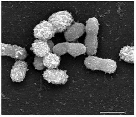
Akkermansia muciniphila
Akkermansia muciniphila는 2004년 사람의 분변에서 분리된 장내 미생물이다. 이 미생물은 장의 상피세포에서 생성되는 뮤신 (mucin)을 분해하여 질소원과 탄소원으로 이용하며 이 과정에서 아세트산 (acetate), 프로피온산 (propionate), 에탄올 (etanol) 등을 부가적으로 생성한다1. A. muciniphila는 사람의 장내에서 발견되는 문(Phylum) 중에 하나인 Verrucomicrobia 문에 속하며, 동물과 사람에게서 건강 연관성이 밝혀져있는 상당한 관심을 받고있는 미생물이다2.
A. muciniphila 는 신생아의 장내에서도 발견되는데, 핀란드의 투르쿠 대학 연구팀에 연구에서 신생아 장내 미생물 군집에서 A. muciniphila 의 풍부도는 생후 1년 안에 건강한 성인의 장에서 관찰되는 수준까지 증가하며 성인에서 노인으로 연령 증가에 따라서 감소하는 경향이 확인되었다3,4. 또한, 염증성 장질환(Inflammatory bowel disease, IBD) 환자와 비만을 포함한 대사 장애 (Metabolic disorder)를 겪고 있는 사람의 장에서 풍부도가 감소하는 경향이 나타나 A. muciniphila가 잠재적으로 항 염증 특성을 갖고 있음이 제시되었다5–7.

다음 표는 임상 연구에서 Akkermansia muciniphila의 풍부도를 관찰한 결과이다.
| 대상 그룹 | 그룹 인원 수 | 분석 방법 | Akkermansia 풍부도 | 논문 |
| 비만 여성 | 비만 여성 53명 | Shotgun metagenomic | 인슐린 저항성과 이상지질혈증 마커와 음의 상관관계 | [8] |
| 우수한 운동선수 | 높은 BM의 우수한 운동선수 40명, 낮은 BMI 건강한 남성 23명, 높은 BMI 건강한 남성 23명 | 16S sequencing | 운동선수와 낮은 BMI의 남성에게서 풍부도가 높음 | [9] |
| 정상, 과체중, 비만 성인 | 정상 10명, 과체중 10명, 비만 10명 | 16S sequencing | BMI가 낮은 사람일수록 풍부도가 높음 | [10] |
| 4-5세 정상, 과체중, 비만 아이 | 정상 20명, 과체중과 비만 20명 | q-PCR, T-RELP | 과체중, 비만 아이에게서 감소 | [7] |
| 비만 여성 | 비만 여성 7명 | 16S sequencing | 체중 감소와 양의 상관관계 | [11] |
| 성인 여성 | 정상 체중 17명, 비만 50명 | q-PCR | 정상 여성에서 풍부도 증가하는 경향 | [5] |
References
- 1.Derrien M. Akkermansia muciniphila gen. nov., sp. nov., a human intestinal mucin-degrading bacterium. INTERNATIONAL JOURNAL OF SYSTEMATIC AND EVOLUTIONARY MICROBIOLOGY. September 2004:1469-1476. doi:10.1099/ijs.0.02873-0
- 2.Derrien M, Belzer C, de Vos WM. Akkermansia muciniphila and its role in regulating host functions. Microbial Pathogenesis. May 2017:171-181. doi:10.1016/j.micpath.2016.02.005
- 3.Collado MC, Derrien M, Isolauri E, de Vos WM, Salminen S. Intestinal Integrity and Akkermansia muciniphila, a Mucin-Degrading Member of the Intestinal Microbiota Present in Infants, Adults, and the Elderly. Applied and Environmental Microbiology. October 2007:7767-7770. doi:10.1128/aem.01477-07
- 4.Cheng J, Ringel-Kulka T, Heikamp-de Jong I, et al. Discordant temporal development of bacterial phyla and the emergence of core in the fecal microbiota of young children. ISME J. October 2015:1002-1014. doi:10.1038/ismej.2015.177
- 5.F.S.Teixeira T, Grześkowiak ŁM, Salminen S, Laitinen K, Bressan J, Gouveia Peluzio M do C. Faecal levels of Bifidobacterium and Clostridium coccoides but not plasma lipopolysaccharide are inversely related to insulin and HOMA index in women. Clinical Nutrition. December 2013:1017-1022. doi:10.1016/j.clnu.2013.02.008
- 6.Png CW, Lindén SK, Gilshenan KS, et al. Mucolytic Bacteria With Increased Prevalence in IBD Mucosa Augment In Vitro Utilization of Mucin by Other Bacteria. American Journal of Gastroenterology. November 2010:2420-2428. doi:10.1038/ajg.2010.281
- 7.Karlsson CLJ, Önnerfält J, Xu J, Molin G, Ahrné S, Thorngren-Jerneck K. The Microbiota of the Gut in Preschool Children With Normal and Excessive Body Weight. Obesity. November 2012:2257-2261. doi:10.1038/oby.2012.110
- 8.Brahe LK, Le Chatelier E, Prifti E, et al. Specific gut microbiota features and metabolic markers in postmenopausal women with obesity. Nutr & Diabetes. June 2015:e159-e159. doi:10.1038/nutd.2015.9
- 9.Clarke SF, Murphy EF, O’Sullivan O, et al. Exercise and associated dietary extremes impact on gut microbial diversity. Gut. June 2014:1913-1920. doi:10.1136/gutjnl-2013-306541
- 10.Escobar JS, Klotz B, Valdes BE, Agudelo GM. The gut microbiota of Colombians differs from that of Americans, Europeans and Asians. BMC Microbiol. December 2014. doi:10.1186/s12866-014-0311-6
- 11.Kim B-S, Song M, Kim H. The anti-obesity effect of Ephedra sinica through modulation of gut microbiota in obese Korean women. Journal of Ethnopharmacology. March 2014:532-539. doi:10.1016/j.jep.2014.01.038

